


Introduction
Nonmelanoma skin cancer (NMSC), including basal cell carcinoma (BCC) and squamous cell carcinoma (SCC), is the most common skin cancer in non-Hispanic whites. Although NMSC-related deaths are rare, more than 1 million new NMSC cases are diagnosed annually in the United States, a number approximately equivalent to the total new cases of all other cancers combined (Jemal et al., 2006). NMSC is found primarily on sun-exposed sites, and a fair complexion and frequent exposure to sunlight are the primary risk factors (Armstrong et al., 1997; English et al., 1997; Strom and Yamamura, 1997; Armstrong and Kricker, 2001; Diepgen and Mahler, 2002). Another possible risk factor is suboptimal cellular DNA repair capacity, because patients with xeroderma pigmentosum have inherited defects in DNA repair and have a more-than-1,000-fold increased risk of sunlight-induced skin cancer (Kraemer et al., 1984; Kraemer et al., 1994; Lambert et al., 1995; Sarasin and Stary, 1997; Norgauer et al., 2003). This suggests that suboptimal DNA repair capacity could be a risk factor for skin cancer in the general population (Wei et al., 1993, 1994a, 1994b; Matta et al., 2003).
One key to the prevention of skin cancer is early identification of at-risk individuals and early detection of the disease. In that effort, biomarkers for genetic susceptibility to skin cancer are vital, but currently few biomarkers are available to serve this purpose. The mutagen sensitivity assay, which was developed by Dr T.C. Hsu (Cherry and Hsu, 1983), measures the number of mutagen-induced chromatid breaks per cell (b/c) in cultured primary peripheral blood lymphocytes. With bleomycin, a radiomimetic chemical, as a test mutagen, Dr Hsu and co-workers successfully used the assay to investigate genetic susceptibility to tobacco-related cancers (Cherry and Hsu, 1983; Spitz et al., 1989, 1995; Fireman et al., 1994; Cloos et al., 1996; Ankathil et al., 1999). Similarly, other test mutagens, such as 4-nitroquinoline-1-oxide (4-NQO), an UV-mimetic chemical (Nagao and Sugimura, 1976) that can cause bulky DNA adducts and chromosomal aberrations in exposed cells (Darroudi et al., 1989), are also used to determine genetic susceptibility to various types of cancer (Hsu et al., 1993a, 1993b; Gu et al., 1999).
In an early pilot study, Hsu et al. (1993a, 1993b) found that lymphocytes from melanoma patients were more susceptible to 4-NQO than those from head and neck cancer patients or healthy subjects. Therefore, we thought that in vitro 4-NQO-induced chromatid breaks in blood lymphocytes could also be used as a biomarker for studying genetic susceptibility to cutaneous malignancies with much simplified experimental procedures compared with those of the assay that uses UV as a mutagen. Since 4-NQO is a UV-mimetic and adduct-forming carcinogen that causes DNA damage uniquely repaired by global genome repair (Snyderwine and Bohr, 1992), it has been widely used in experimental research for cellular responses to DNA damage (Kyng et al., 2005), DNA repair (Ninomiya et al., 2004), and induced mutation spectrum in genomic (Muftuoglu et al., 2002) and mitochondrial DNA (Mambo et al., 2003).
However, no published studies, to our knowledge, have established an association between 4-NQO-induced mutagen sensitivity and risk of skin cancer in the general population. To further explore the feasibility of using 4-NQO as a mutagen substitute for UV and its relevance to host DNA repair capacity in lymphocytes, and to extend our earlier findings in the pilot study (Wei et al., 1996a), we tested our hypothesis in a larger, independent hospital-based case–control study of sporadic NMSC that 4-NQO-induced mutagen sensitivity is associated with the development of NMSC in the general population.
Results
Characteristics of study subjects
We first examined the differences in the distributions of
age, sex, and selected risk factors between the cases and the controls. The
analysis included 191 cases (110 BCC and 81 SCC) and 176 cancer-free controls.
All subjects were non-Hispanic whites. The distributions of age, sex, and
selected risk factors for the cases and controls are presented in Table
1. The age range (mean.gif) SD)
was 23–85 (59.014.1)
years for BCC cases, 34–93 (65.512.2)
years for SCC cases, and 23–83 (55.513.5)
years for the controls. However, patients with BCC and SCC tended to be older
than the controls. The proportion of men was higher in patients with BCC (62.7%)
and SCC (88.9%) than in the controls (46.0%; Table
1). Since these differences could be due to possible biases in selecting the
controls in a hospital setting, they were adjusted for in the following
multivariate logistic regression analyses.
SD)
was 23–85 (59.014.1)
years for BCC cases, 34–93 (65.512.2)
years for SCC cases, and 23–83 (55.513.5)
years for the controls. However, patients with BCC and SCC tended to be older
than the controls. The proportion of men was higher in patients with BCC (62.7%)
and SCC (88.9%) than in the controls (46.0%; Table
1). Since these differences could be due to possible biases in selecting the
controls in a hospital setting, they were adjusted for in the following
multivariate logistic regression analyses.
Table 1 - Distribution of selected risk factors between patients with nonmelanoma skin cancer and controls and logistic regression analysis.
 Full
table
Full
table
Risk factors for NMSC
Using a multivariate logistic regression analysis with adjustment for age and sex, we assessed the odds ratios (ORs) and 95% confidence intervals (CIs) to estimate the risk of NMSC associated with known risk factors, such as hair color, eye color, skin color, tanning ability, number of sunburns, freckling, dysplastic nevi, and family history of skin cancer. It was apparent that these risk factors were associated with a significantly increased risk for BCC (except for eye color and dysplastic nevi) and SCC (except for hair color, eye color, and dysplastic nevi) in this study population (Table 1). For example, the most pronounced risk factors were freckling (OR=3.71, 95% CI (2.13–6.44)) and family history of skin cancer (OR=2.94, 95% CI (1.63--5.28)) for BCC and one or more sunburns (OR=3.36, 95% CI (1.68–6.72)) and family history of skin cancer (OR=2.98, 95% CI (1.40–6.36)) for SCC. These risk factors were also correlated with each other, as expected. For example, hair color was statistically significantly correlated with eye color (r=0.218, P=0.004), skin color (r=0.223, P=0.003), tanning ability (r=0.210, P=0.006), number of sunburns (r=0.191, P=0.012), and freckling in the sun (r=0.216, P=0.005) in the controls (data not shown). Overall, the data suggested that these risk factors played a role in sporadic NMSCs in this study population.
4-NQO-induced chromatid breaks
We then examined the difference in the frequencies of
4-NQO-induced chromatid b/c between the cases and the controls. The number of
simple chromatid breaks was scored from 50 well-spread metaphases for each
subject and expressed as the number of chromatid b/c, as Lee
et al. (1996) showed that the statistical efficiency of reading 50,
compared with 100 metaphases was similar. Because the mean spontaneous b/c value
derived from 50 metaphases of untreated cells was 0.02 (Wei
et al., 1996b; Wang
et al., 2000), which was more-than-10-fold less than that of 4-NQO
treated cells (0.25 b/c in the controls), we used 4-NQO-induced b/c values only
for statistical comparisons, as recommended by Hsu
et al. (1989). Since the results of both log-transformed and
untransformed data were very similar (data not shown), we presented the results
of the untransformed data only for simplicity. The percentage distributions of
4-NQO-induced b/c values in BCC cases, SCC cases, and the controls are shown in
Figure
1. It was apparent that the distributions of 4-NQO-induced b/c values in
both BCC and SCC cases tended to be higher values than those of the controls. We
did not perform a separate analysis of patients with a history of NMSC, because
there was no statistical difference in b/c values between cases with and without
previous NMSC (52 and 58 BCC cases, respectively, with meanSD,
0.390.34
vs 0.470.42
(P=0.322); 33 and 48 SCC cases, respectively, with 0.540.55
vs 0.400.32
(P=0.204)). The 4-NQO-induced b/c values were statistically higher in
both BCC and SCC cases than in the controls (P<0.001 for both
comparisons; Table
2).
Figure 1.
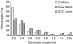
Percentage distributions of chromatid b/c in BCC cases, SCC cases, and controls.
Full figure (14K)Table 2 - Comparison of differences in frequencies of chromatid breaks per cell induced by 4-NQO between patients with nonmelanoma skin cancer and controls.
Full
table
After stratification by age, sex, and selected risk factors, the
mean 4-NQO-induced b/c values remained statistically significantly higher in
both BCC and SCC cases than in the controls for each subgroup
(P<0.05), except for dysplastic nevi (P=0.457) for BCC and age
younger than 55 years (P=0.123), blond or red hair (P=0.482), fair
skin (P=0.073), and family history of skin cancer (P=0.082) for
SCC. We suspected that these anomalies might be resulted from the smaller
numbers of observations in the strata (Table
2). We also compared the differences in the strata in each of the subgroups,
and found that only SCC patients with freckling in the sun as a child were
borderline significantly sensitive to 4-NQO, as indicated by higher frequencies
of chromatid breaks than SCC patients without freckling (P=0.053).
Finally, we evaluated whether the sensitivity to 4-NQO was associated with tumor
behavior, but we did not find any difference in 4-NQO-induced b/c values between
93 nonaggressive and 17 aggressive cases of BCC (meanSD,
0.430.39
vs 0.420.36,
P=0.922) or between 48 nonaggressive and 33 aggressive cases of SCC
(0.460.39
vs 0.460.49,
P>0.99). Therefore, we did not consider nonaggressive and aggressive
tumors separately in the following analyses.
Association between 4-NQO-induced b/c values and risk of NMSC
We next performed multivariate logistic regression analysis to calculate ORs and 95% CIs with adjustment for only age and sex to avoid attrition due to missing data for selected risk factors (Table 3). The 4-NQO-induced b/c values were fitted in the logistic regression model either as continuous or categorical variables. We found that the risk associated with an increment of 0.1 in the b/c value was 1.22 (95% CI (1.12–1.34)) for BCC and 1.26 (95% CI (1.12–1.41)) for SCC after adjustment for age and sex (Table 3). Using the median b/c value of the controls as the cutoff point, high b/c values were associated with a more-than-twofold increased risk for both BCC (OR=2.69, 95% CI (1.59–4.54)) and SCC (OR=2.46, 95% CI (1.28–4.71)) after adjustment for age and sex (Table 3). We also used 4-NQO-induced b/c tertile values of the controls as cutoff points to further evaluate the trend of the effects. We found that as the b/c values increased, the ORs increased for both BCC and SCC. Specifically, compared with the lower tertile, higher b/c values were associated with increased risk in a dose-dependent manner for both BCC (OR=1.50, 95% CI (0.72–3.09) for the middle tertile; OR=4.04, 95% CI (2.08–7.85) for the upper tertile; and SCC (OR=1.66, 95% CI (0.67–4.13) for the middle tertile; OR=4.72, 95% CI (2.07–10.75) for the upper tertile; Ptrend <0.001 for both BCC and SCC; Table 3).
Table 3 - Logistic regression analysis of chromatid breaks per cell in patients with nonmelanoma skin cancer and cancer-free controls.
Full
table
Multivariate analysis
Afterwards, we included all the selected risk factors in the multivariate logistic regression model for those who had provided complete information in a smaller data set (89 BCC cases, 67 SCC cases, and 126 controls). Using simultaneous adjustment for all variables listed in Table 4, we found that statistically significantly increased risk was associated with both age and sex in both BCC and SCC, but these could be adjusted as confounders due to possible selection bias in the hospital-based study. Most of the known sun-exposure-related risk factors were not significant in this model. It is likely that they were overadjusted by inclusion in the same model due to the correlation among them or limited sample size that may have caused reduction in study power. For example, lifetime sunburns was associated with a borderline significantly 2.5-fold increased risk for SCC but not for BCC (OR=1.35), whereas freckling was associated with 2.3-fold increased risk for BCC (OR=2.27, 95% CI (1.12–4.60); Table 4) but not for SCC (OR=1.45). However, family history of skin cancer remained a significant risk factor for both BCC (OR=2.75, 95% CI (1.29–5.87)) and SCC (OR=3.07, 95% CI (1.11–8.50)). More importantly, independent of these variables, risks associated with 4-NQO-induced b/c values greater than the control median remained higher for both BCC (OR=3.69, 95% CI (1.89–7.19)) and SCC (OR=4.02, 95% CI (1.68–9.60); Table 4). Therefore, these data suggested that in vitro sensitivity to 4-NQO was an independent risk factor for both BCC and SCC in this study population.
Table 4 - Multivariate logistic regression analysis of associations between the frequency of 4-NQO-induced chromosome breaks and risk for BCC and SCC.
Full
table
Interactions between 4-NQO-induced b/c and selected risk factors
We then assessed possible interactions between 4-NQO-induced chromatid breaks and each of the selected risk factors in multivariate logistic regression models. The hypotheses of additive and multiplicative interactions were tested when we included the interaction (or cross-product) terms (i.e., dichotomized mutagen sensitivity X each dichotomized risk factor) in a multivariate logistic regression model that included age, sex, main effect of mutagen sensitivity, and corresponding other risk factors. A departure from the multiplicative model was indicated if the odds ratio for the interactive term was greater than 1; however, there was no evidence of multiplicative interactions between mutagen sensitivity and any of selected risk factors. All hypotheses that failed to reject a multiplicative model were further tested in additive models, as assessed by the 95% confidence bounds. The following pairs of variables that were beyond the bounds suggested a possibility of a more-than-additive effect: freckling in the sun as a child and mutagen sensitivity (the P-value for the additive interactions was 0.002), tanning ability after prolonged sun exposure and mutagen sensitivity (pinteraction=0.026), and family history of skin cancer and mutagen sensitivity (pinteraction=0.049) for BCC, but not for SCC (data not shown).
Correlation between 4-NQO- and UV-induced b/c
For the subjects tested for 4-NQO sensitivity, their sensitivity to UV-B was also tested, and the data were published in a separate study (Wang et al., 2005). As 4-NQO is considered a UV-mimetic agent, we assessed the correlation between these two sets of mutagen sensitivity data as shown in Figure 2. There were 157 cancer-free control subjects whose data on both UV-induced b/c and 4-NQO-induced b/c values were available. However, we did not find evidence for a correlation between these two data sets (Figure 2).
Figure 2.
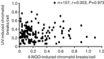
Correlation between 4-NQO- and UV-induced chromatid b/c in cancer-free controls.
Full figure (13K)Discussion
In this case–control study, we found that sunlight-related risk factors played a role in both BCC and SCC, and that 4-NQO-induced mutagen sensitivity was an independent risk factor for both BCC and SCC in a dose–response manner in this study population. NMSC patients had significantly higher frequencies of 4-NQO-induced chromatid breaks that were associated with more-than-twofold increased risk for NMSC compared with the cancer-free controls. Therefore, a mutagen sensitivity assay using 4-NQO as the test mutagen may be used to assess genetic susceptibility to NMSC in the general population.
Although mutagen sensitivity was measured in cultured peripheral lymphocytes, high susceptibility to carcinogen-induced chromatid breaks has a clear genetic basis (Cloos et al., 1999; Tedeschi et al., 2004), and either spontaneous or in vitro-induced chromosomal aberrations by mutagens or carcinogens are relevant to cutaneous carcinogenesis in the target organ (Wei et al., 1996a; Lanza et al., 1997). The feasibility of procuring peripheral blood samples, the ease of using this in vitro 4-NQO mutagen sensitivity assay, and the small amount (2 ml) of blood needed for this assay make it a promising biomarker for assessing individual susceptibility to cancer. However, the usefulness of this assay in assessing susceptibility to cancers other than NMSC in the general population needs further investigation.
We previously published the data on UV-B-induced mutagen sensitivity and risk of NMSC in the same study population, in which we found that UV-B-induced chromatid breaks were associated with risk of developing BCC and SCC (Wang et al., 2005). However, we did not find the correlation between UV-B-induced b/c and 4-NQO-induced b/c in 157 control subjects who had been assessed by both of the assays. It is suggested that even though 4-NQO is similar to UV light in the molecular mechanisms of inducing mutagenesis such as base change, deletion, or phage induction ability, and repair in xeroderma pigmentosum fibroblastic cells such as DNA synthesis or colony-forming ability (Nagao and Sugimura, 1976), the formation of chromatid breaks induced by these two agents may have different mechanisms, especially in repair-proficient subjects. It is known that the UV-induced DNA lesions are mainly repaired by the transcription-coupled repair pathway, whereas the lesions induced by 4-NQO are repaired by the global genome repair pathway (Snyderwine and Bohr, 1992; Muftuoglu et al., 2002). However, no previously published report has investigated the correlation between damage to DNA and chromosomes induced by these two carcinogens in the general population. Therefore, our results remain to be validated by other investigators.
Although the exact mechanisms by which UV and 4-NQO induce chromosomal breaks are unknown, it is well known that during the repair of the lesions, repair enzymes can make more cuts on the damaged DNA strands (Marcon and Moens, 2005), which can cause double-strand breaks. A slow kinetics and suboptimal synapsis mechanism of back-up non-homologous end-joining of the double-strand breaks has been proposed (Iliakis et al., 2004), in which more time needed for exchanges through the joining of incorrect ends may cause the formation of chromosome aberrations. However, the mechanisms by which these lesions are converted into chromosome breaks are still poorly understood (Slijepcevic and Bryant, 1998). One of the mechanisms may be that the conversion is mediated by the enzyme telomerase, a possible mechanism called telomere capture that is intended to stabilize broken chromosomes in mammalian cells (Meltzer et al., 1993; Bosco and Haber, 1998).
Recent evidence also suggests that chromatid breaks are not simply the expanded DNA double-strand breaks, and that there is a lack of correlation between reduced DNA double-strand break rejoining and disappearance of chromatid breaks. As a result, a modified Revell's model has been proposed that in less than 20% of the total breaks, both spontaneous and radiation-induced chromatid breaks result from DNA double-strand breaks signaling and rearrangement processes from within large looped chromatin domains (Bryant, 1998), and that the rest may result from intrachromatid rearrangements, including a very small proportion involving complete excision of a looped domain (Bryant et al., 2004). Nevertheless, whether different DNA repair mechanisms are involved in the formation of the UV- and 4-NQO-induced chromatid breaks remains to be determined.
There were some observable differences in the sensitivity to in vitro exposure to 4-NQO not only between BCC and SCC but also within each cancer site. For example, an apparently higher level of 4-NQO-induced chromatid breaks was observed in young BCC but in old SCC patients, in BCC patients with blond or red hair but in SCC patients with black or brown hair. This may reflect the fact that genetic susceptibility (i.e., early age onset or having some genetic traits) plays a major role in BCC, whereas overexposure to sunlight (i.e., accumulative dose characterized by older age and no genetic traits) is the major cause for SCC (Armstrong and Kricker 2001). However, there were also some inconsistencies in the findings. For example, an apparently higher level of 4-NQO-induced chromatid breaks was also observed in both BCC and SCC patients who had no history of sunburns, had freckling, or had not reported a family history of skin cancer. These results may also reflect instability of the estimates, because the sample size of a given subgroup or stratum was substantially reduced in the stratification, suggesting a larger study may be needed to verify these findings.
In summary, we found that the frequency of 4-NQO-induced chromatid breaks was significantly higher in NMSC cases than in the controls. This higher frequency was associated with a more-than-twofold increased risk for both BCC and SCC, and a dose–response relationship was found between the tertile of mutagen sensitivity and the risk for both BCC and SCC, independent of other known risk factors. These findings suggest that in vitro 4-NQO-induced mutagen sensitivity does not appear to be correlated to that induced by UV, which also reflects susceptibility to NMSC. However, we did not have accumulative sun exposure data for each subject, which may have an effect on DNA repair phenotype. However, the 4-NQO-induced frequency of chromatid breaks values was 10 times higher than that in the untreated cells, suggesting that sun-induced chromatid breaks in lymphocytes may be negligible. Furthermore, we used the in vitro treatment with the same conditions for cases and controls and adjusted possible sun-exposure confounders (sunburn, freckling, and tanning ability) in the multivariate logistic models. Therefore, the sunlight-induced chromatid breaks should not have, if any, a major effect on the b/c values we used for comparison. Finally, because of the inherent bias in selecting study subjects in a hospital-based case–control study, such as potential selection bias in sunlight exposure and self-reported skin lesions, and the relatively small sample size of this study, the findings need further validation by other independent, larger or prospective studies.
Materials and Methods
Study population
The study included patients with BCC and SCC who registered at the Departments of Dermatology and Head and Neck Surgery at The University of Texas M.D. Anderson Cancer Center and DermSurgery Associates from July 1996 to August 2000. There were no restrictions on age, sex, or ethnicity. Controls were reportedly cancer-free healthy people selected from genetically unrelated visitors who were accompanying cancer patients to the clinics at M.D. Anderson Cancer Center or came to M.D. Anderson for prostate cancer screening. The exclusion criteria were previous chemotherapy or radiotherapy, any metastasis, and any cancer history other than NMSC for case subjects and any blood transfusion in the last 6 months for all participants. Informed consent was obtained from all participants, and a standardized, self-administered questionnaire was used to collect demographic data and data on risk factors, such as natural hair color, eye color, skin color, history of sunlight exposure (including freckling in the sun as a child, tanning ability, and number of sunburns), medical history, and family history of first-degree relatives with any cancer. Each participant donated 20 ml of blood, which was collected with heparinized tubes. This analysis included only non-Hispanic whites. The study was conducted according to the Declaration of Helsinki Principles, and the study protocol was approved by the institutional review board of M.D. Anderson.
Mutagen sensitivity assay
In this assay, mutagen sensitivity was expressed as the
number of 4-NQO-induced chromatid b/c after 24 hours of treatment (Hsu
et al., 1993a). Briefly, short-term cultures of 1 ml of fresh
whole blood were established in 9 ml of RPMI 1640 medium supplemented with
20% fetal bovine serum with a final concentration of 112.5 .gif) g/ml
phytohemagglutinin (Remel, Lenexa, KS) to stimulate T-lymphocyte growth. After
48 hours of culture, the cells were treated with 4-NQO at a final
concentration of 10 M and were allowed to grow for another 23 hours before
being treated with colcemid (Gibco BRL, Carlsbad, CA) at 0.06 g/ml
to induce mitotic arrest 1 hour before harvesting. We used conventional
chromosome harvesting procedures: the cells were treated for 15 minutes
with 60 mM hypotonic KCl solution and fixed three
times for 5 minutes, each with freshly prepared methanol:acetic acid (3:1
v/v), after which air-dried slides were prepared as previously described (Cherry
and Hsu, 1983). All the slide preparation procedures were performed by one
of us (Ping Xiong). The slides were then stained with 4% Giemsa (Biomedical
Specialties, Santa Monica, CA) for 7 minutes. All slides were evaluated for
chromosomal aberrations by the late Dr Hsu, who was masked to the case–control
status of the subjects. The number of simple chromatid breaks was scored from 50
well-spread metaphases per blood sample.
g/ml
phytohemagglutinin (Remel, Lenexa, KS) to stimulate T-lymphocyte growth. After
48 hours of culture, the cells were treated with 4-NQO at a final
concentration of 10 M and were allowed to grow for another 23 hours before
being treated with colcemid (Gibco BRL, Carlsbad, CA) at 0.06 g/ml
to induce mitotic arrest 1 hour before harvesting. We used conventional
chromosome harvesting procedures: the cells were treated for 15 minutes
with 60 mM hypotonic KCl solution and fixed three
times for 5 minutes, each with freshly prepared methanol:acetic acid (3:1
v/v), after which air-dried slides were prepared as previously described (Cherry
and Hsu, 1983). All the slide preparation procedures were performed by one
of us (Ping Xiong). The slides were then stained with 4% Giemsa (Biomedical
Specialties, Santa Monica, CA) for 7 minutes. All slides were evaluated for
chromosomal aberrations by the late Dr Hsu, who was masked to the case–control
status of the subjects. The number of simple chromatid breaks was scored from 50
well-spread metaphases per blood sample.
Statistical analysis
The number of chromatid b/c was analyzed as a continuous
variable. Student's t-test was used to compare the mean number of
chromatid b/c between groups. As the chromatid b/c were not normally
distributed, we also performed Student's t-test for log-transformed data.
We used the median and tertile of chromatid b/c in the control group as the
cutoff values to calculate the OR. Correlation analysis was used to explore the
relationships between sun exposure variables. Univariate and multivariate
logistic regression analyses with adjustment for age and sex were performed to
calculate the adjusted OR and 95% CI for each variable of interest. Some
subjects did not provide information on some variables (such as hair color, eye
color, skin color, tanning ability, number of sunburns, freckling, dysplastic
nevi, and family history of skin cancer), and these variables were treated as
missing data in the analysis. Afterward, we included all the selected variables
in the multivariate logistic regression analyses for subjects who provided
complete information. A more-than-multiplicative interaction was suggested when
OR11>OR10 .gif) OR01, in which OR11=the OR when both factors were present,
OR01=the OR when only factor 1 was present and OR10=the OR
when only factor 2 was present (Kleinbaum
et al., 1982). To assess evidence for departure from a multiplicative
model, we modeled interaction terms between variables using standard
unconditional logistic regression. We were specifically interested in searching
for interactions indicating a more-than-multiplicative relationship (i.e.,
interaction terms from the logistic regression with positive coefficients),
because these interactions identify subgroups of individuals who may be at
particularly high risk for developing NMSC. We were also interested in
identifying departures from additive models. Empirically, a more-than-additive
interaction was indicated if
OR11>OR10+OR01-1. When the test for
multiplicative interaction was not rejected, further tests for additive
interaction were performed by a bootstrapping test of goodness-of-fit of the
null hypothesis of an additive model with no interaction against an alternative
hypothesis that allows an additive interaction. To perform the hypothesis test
for additive models, we implemented bootstrapping using Stata 8.2 (StataCorp LP,
College Station, TX). All statistical tests were two-sided, and P<0.05
was considered statistically significant. We analyzed all data, except for
additive models, using SAS software (version 8e; SAS Institute, Cary, NC).
OR01, in which OR11=the OR when both factors were present,
OR01=the OR when only factor 1 was present and OR10=the OR
when only factor 2 was present (Kleinbaum
et al., 1982). To assess evidence for departure from a multiplicative
model, we modeled interaction terms between variables using standard
unconditional logistic regression. We were specifically interested in searching
for interactions indicating a more-than-multiplicative relationship (i.e.,
interaction terms from the logistic regression with positive coefficients),
because these interactions identify subgroups of individuals who may be at
particularly high risk for developing NMSC. We were also interested in
identifying departures from additive models. Empirically, a more-than-additive
interaction was indicated if
OR11>OR10+OR01-1. When the test for
multiplicative interaction was not rejected, further tests for additive
interaction were performed by a bootstrapping test of goodness-of-fit of the
null hypothesis of an additive model with no interaction against an alternative
hypothesis that allows an additive interaction. To perform the hypothesis test
for additive models, we implemented bootstrapping using Stata 8.2 (StataCorp LP,
College Station, TX). All statistical tests were two-sided, and P<0.05
was considered statistically significant. We analyzed all data, except for
additive models, using SAS software (version 8e; SAS Institute, Cary, NC).
References
- Ankathil R, Jyothish B, Madhavan J, Nair MK (1999) Deficient DNA repair capacity: a predisposing factor and high risk predictive marker in familial colorectal cancer. J Exp Clin Cancer Res 18:33–37 | PubMed | ChemPort |
- Armstrong BK, Kricker A (2001) The epidemiology of UV induced skin cancer. J Photochem Photobiol B 63:8–18 | Article | PubMed | ISI | ChemPort |
- Armstrong BK, Kricker A, English DR (1997) Sun exposure and skin cancer. Austral J Dermatol 38(Suppl 1):S1–S6
- Bosco G, Haber JE (1998) Chromosome break-induced DNA replication leads to nonreciprocal translocations and telomere capture. Genetics 150:1037–1047 | PubMed | ISI | ChemPort |
- Bryant PE (1998) The signal model: a possible explanation for the conversion of DNA double-strand breaks into chromatid breaks. Int J Radiat Biol 73:243–251 | Article | PubMed | ChemPort |
- Bryant PE, Gray LJ, Peresse N (2004) Progress towards understanding the nature of chromatid breakage. Cytogenet Genome Res 104:65–71 | Article | PubMed | ChemPort |
- Cherry LM, Hsu TC (1983) Bleomycin-induced chromosome damage in lymphocytes of medullary thyroid carcinoma patients and their family members. Anticancer Res 3:367–372 | PubMed | ISI | ChemPort |
- Cloos J, Nieuwenhuis EJ, Boomsma DI, Kuik DJ, van der Sterre ML, Arwert F et al. (1999) Inherited susceptibility to bleomycin-induced chromatid breaks in cultured peripheral blood lymphocytes. J Natl Cancer Inst 91:1125–1130 | Article | PubMed | ChemPort |
- Cloos J, Spitz MR, Schantz SP, Hsu TC, Zhang ZF, Tobi H et al. (1996) Genetic susceptibility to head and neck squamous cell carcinoma. J Natl Cancer Inst 88:530–535 | PubMed | ChemPort |
- Darroudi F, Natarajan AT, Lohman PH (1989) Cytogenetical characterization of UV-sensitive repair-deficient CHO cell line 43-3B. II. Induction of cell killing, chromosomal aberrations and sister-chromatid exchanges by 4NQO, mono- and bi-functional alkylating agents. Mutat Res 212:103–112 | PubMed | ChemPort |
- Diepgen TL, Mahler V (2002) The epidemiology of skin cancer. Br J Dermatol 146(Suppl 61):1–6 | Article | PubMed | ISI |
- English DR, Armstrong BK, Kricker A, Fleming C (1997) Sunlight and cancer. Cancer Causes Control 8:271–283 | Article | PubMed | ISI | ChemPort |
- Fireman Z, Shabtai F, Lurie B (1994) Chromosome sensitivity to bleomycin-induced mutagenesis in lymphocytes from colorectal cancer patients under 40 years of age. Dis Colon Rectum 37:1317–1320 | Article | PubMed | ChemPort |
- Gu J, Bondy ML, Sigurdson A, Spitz MR, Hsu TC, Wu X (1999) Three measures of mutagen sensitivity in a cancer-free population. Cancer Genet Cytogenet 110:65–69 | Article | PubMed | ChemPort |
- Hsu TC, Feun L, Trizna Z, Savaraj N, Shirley LR, Furlong CL et al. (1993b) Differential sensitivity among three human subpopulations in response to 4-nitroquinoline-1-oxide and to bleomycin. Int J Oncol 3:827–830 | ChemPort |
- Hsu TC, Johnston DA, Cherry LM, Ramkissoon D, Schantz SP, Jessup JM et al. (1989) Sensitivity to genotoxic effects of bleomycin in humans: possible relationship to environmental carcinogenesis. Int J Cancer 43:403–409 | PubMed | ISI | ChemPort |
- Hsu TC, Savaraj N, Trizna Z, Feun L, Furlong CL, Schantz SP et al. (1993a) Cytogenetic studies on the in vitro genotoxicity of 4-nitroquinoline-1-oxide on human lymphocytes. Int J Oncol 3:823–826 | ChemPort |
- Jemal A, Siegel R, Ward E, Murray T, Xu J, Smigal C et al. (2006) Cancer statistics, 2006. CA Cancer J Clin 56:106–130 | PubMed |
- Iliakis G, Wang H, Perrault AR, Boecker W, Rosidi B, Windhofer F et al. (2004) Mechanisms of DNA double strand break repair and chromosome aberration formation. Cytogenet Genome Res 104:14–20 | Article | PubMed | ChemPort |
- Kleinbaum D, Kupper L, Morgenstern H (1982) Epidemiologic research: principles and quantitative methods. Lifetime Learning: London, 529
- Kraemer KH, Lee MM, Andrews AD, Lambert WC (1994) The role of sunlight and DNA repair in melanoma and nonmelanoma skin cancer. The xeroderma pigmentosum paradigm. Arch Dermatol 130:1018–1021 | Article | PubMed | ISI | ChemPort |
- Kraemer KH, Lee MM, Scotto J (1984) DNA repair protects against cutaneous and internal neoplasia: evidence from xeroderma pigmentosum. Carcinogenesis 5:511–514 | PubMed | ISI | ChemPort |
- Kyng KJ, May A, Stevnsner T, Becker KG, Kolvra S, Bohr VA (2005) Gene expression responses to DNA damage are altered in human aging and in Werner Syndrome. Oncogene 24:5026–5042 | Article | PubMed | ChemPort |
- Lambert WC, Kuo HR, Lambert MW (1995) Xeroderma pigmentosum. Dermatol Clin 13:169–209 | PubMed | ChemPort |
- Lanza A, Lagomarsini P, Casati A, Ghetti P, Stefanini M (1997) Chromosomal fragility in the cancer-prone disease xeroderma pigmentosum preferentially involves bands relevant for cutaneous carcinogenesis. Int J Cancer 74:654–663 | Article | PubMed | ChemPort |
- Lee JJ, Trizna Z, Hsu TC, Spitz MR, Hong WK (1996) A statistical analysis of the reliability and classification error in application of the mutagen sensitivity assay. Cancer Epidemiol Biomarkers Prev 5:191–197 | PubMed | ChemPort |
- Mambo E, Gao X, Cohen Y, Guo Z, Talalay P, Sidransky D (2003) Electrophile and oxidant damage of mitochondrial DNA leading to rapid evolution of homoplasmic mutations. Proc Natl Acad Sci USA 100:1838–1843 | Article | PubMed | ChemPort |
- Marcon E, Moens PB (2005) The evolution of meiosis: recruitment and modification of somatic DNA-repair proteins. Bioessays 27:795–808 | Article | PubMed | ChemPort |
- Matta JL, Villa JL, Ramos JM, Sanchez J, Chompre G, Ruiz A et al. (2003) DNA repair and nonmelanoma skin cancer in Puerto Rican populations. J Am Acad Dermatol 49:433–439 | Article | PubMed |
- Meltzer PS, Guan X-Y, Trent JM (1993) Telomere capture stabilizes chromosome breakage. Nat Genet 4:252–255 | Article | PubMed | ISI | ChemPort |
- Muftuoglu M, Selzer R, Tuo J, Brosh RM Jr, Bohr VA (2002) Phenotypic consequences of mutations in the conserved motifs of the putative helicase domain of the human Cockayne syndrome group B gene. Gene 283:27–40 | Article | PubMed | ChemPort |
- Nagao M, Sugimura T (1976) Molecular biology of the carcinogen, 4-nitroquinoline 1-oxide. Adv Cancer Res 23:131–169 | PubMed | ChemPort |
- Ninomiya Y, Suzuki K, Ishii C, Inoue H (2004) Highly efficient gene replacements in Neurospora strains deficient for nonhomologous end-joining. Proc Natl Acad Sci USA 101:12248–12253 | Article | PubMed | ChemPort |
- Norgauer J, Idzko M, Panther E, Hellstern O, Herouy Y (2003) Xeroderma pigmentosum. Eur J Dermatol 13:4–9 | PubMed |
- Sarasin A, Stary A (1997) Human cancer and DNA repair-deficient diseases. Cancer Detect Prev 21:406–411 | PubMed | ChemPort |
- Slijepcevic P, Bryant PE (1998) Chromosome healing, telomere capture and mechanisms of radiation-induced chromosome breakage. Int J Radiat Biol 73:1–13 | Article | PubMed | ISI | ChemPort |
- Snyderwine EG, Bohr VA (1992) Gene- and strand-specific damage and repair in Chinese hamster ovary cells treated with 4-nitroquinoline 1-oxide. Cancer Res 52:4183–4189 | PubMed | ChemPort |
- Spitz MR, Fueger JJ, Beddingfield NA, Annegers JF, Hsu TC, Newell GR et al. (1989) Chromosome sensitivity to bleomycin-induced mutagenesis, an independent risk factor for upper aerodigestive tract cancers. Cancer Res 49:4626–4628 | PubMed | ISI | ChemPort |
- Spitz MR, Hsu TC, Wu X, Fueger JJ, Amos CI, Roth JA (1995) Mutagen sensitivity as a biological marker of lung cancer risk in African Americans. Cancer Epidemiol Biomarkers Prev 4:99–103 | PubMed | ChemPort |
- Strom SS, Yamamura Y (1997) Epidemiology of nonmelanoma skin cancer. Clin Plast Surg 24:627–636 | PubMed | ISI | ChemPort |
- Tedeschi B, Cicchetti R, Argentin G, Caporossi D, Pittaluga M, Parisi P et al. (2004) Aphidicolin and bleomycin induced chromosome damage as biomarker of mutagen sensitivity: a twin study. Mutat Res 546:55–64 | PubMed | ChemPort |
- Wang LE, Bondy ML, de Andrade M, Strom SS, Wang X, Sigurdson A et al. (2000) Gender difference in smoking effect on chromosome sensitivity to gamma radiation in a healthy population. Radiat Res 154:20–27 | Article | PubMed | ChemPort |
- Wang LE, Xiong P, Strom SS, Goldberg LH, Lee JE, Ross MI et al. (2005) In vitro sensitivity to ultraviolet B light and skin cancer risk: a case–control analysis. J Natl Cancer Inst 97:1822–1831 | PubMed | ChemPort |
- Wei Q, Gu J, Cheng L, Bondy ML, Jiang H, Hong WK et al. (1996b) Benzo(a)pyrene diol epoxide-induced chromosomal aberrations and risk of lung cancer. Cancer Res 56:3975–3979 | ChemPort |
- Wei Q, Matanoski GM, Farmer ER, Hedayati MA, Grossman L (1993) DNA repair and aging in basal cell carcinoma: a molecular epidemiology study. Proc Natl Acad Sci USA 90:1614–1618 | Article | PubMed | ChemPort |
- Wei Q, Matanoski GM, Farmer ER, Hedayati MA, Grossman L (1994a) DNA repair and susceptibility to skin cancer. Am J Epidemiol 140:598–607 | PubMed | ISI | ChemPort |
- Wei Q, Matanoski GM, Farmer ER, Hedayati MA, Grossman L (1994b) DNA repair related to multiple skin cancers and drug use. Cancer Res 54:437–440 | PubMed | ISI | ChemPort |
- Wei Q, Spitz MR, Gu J, Cheng L, Xu X, Strom SS et al. (1996a) DNA repair capacity correlates with mutagen sensitivity in lymphoblastoid cell lines. Cancer Epidemiol Biomarkers Prev 5:199–204 | ChemPort |
Acknowledgments
This work was supported in part by the National Institutes of Health National Cancer Institute Grants P01 CA 68233 (G.L.C.), R01 CA 100264 (Q.W.), and the National Institute of Environmental Health Sciences Grant R01 ES11740 (Q.W.). We thank Margaret Lung for assistance in recruiting study participants, Yijue Zhao for laboratory assistance, Monica Domingue, and Joanne Sider for assistance in preparing the manuscript, and David Galloway for scientific editing.


